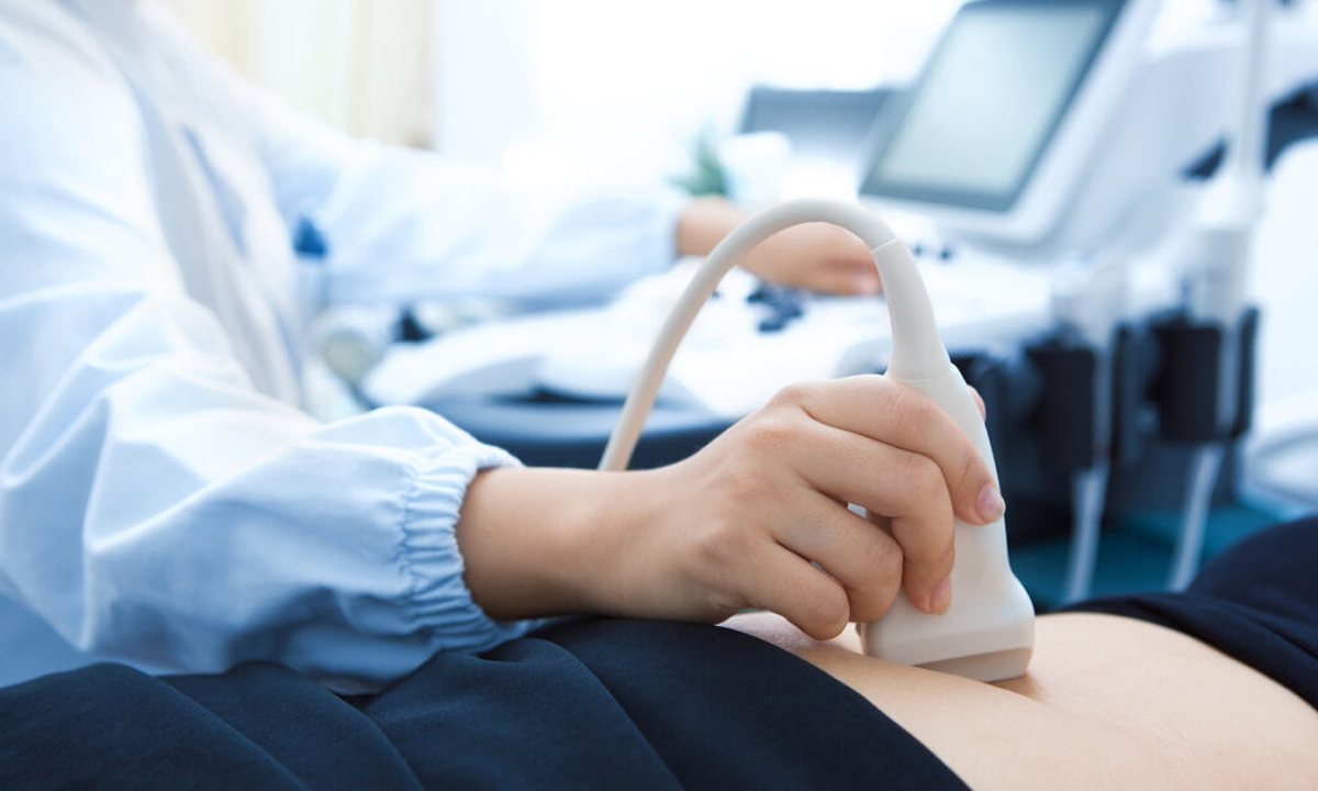
Ultrasound test, also known as USG, sonography test or ultrasonography test, is a non-invasive, painless and radiation free mode of imaging technique used to assess various abdominal and pelvic organs, vascular structure, muscles and tendons. It is also performed to evaluate any congenital abnormalities, well-being and optimal growth of a foetus during pregnancy.
Ultrasound for pregnancy or USG for pregnancy is a primary mode of diagnostic imaging technique to provide diagnosis during pregnancy.
With the advances in ultrasound technology, we can obtain 3D ultrasound and 4D ultrasound images of the foetus, know the architecture of the liver using shear wave technique and demarcate the lesions in thyroid and breast by elastography.

A three-dimensional i.e.3D ultrasound is one of the most advanced techniques of ultrasound in which a standard, two-dimensional greyscale ultrasound image is converted into a volumetric, three-dimensional image.
With the help of post-processing techniques and AI (artificial intelligence), the acquired image is enhanced and can be viewed in all the three-axis for a detailed examination. This technique has been developed in order to solve issues related to obstetrics and gynaecology thereby reducing the dependence on ultrasonography test and also avoid operator related insufficiencies.
A four-dimensional i.e. 4D ultrasound is a real-time, dynamic ultrasound that complements 2D and 3D examination. By use of 4D ultrasound, the medical practitioner can assess dynamic images of the foetal face, respiratory movements, swallowing, movements of the lips and mouth, blinking of eyes and limbs movements.
A 3D ultrasound can be useful in gynaecology for the detection of uterine anomalies such as Mullerian duct abnormalities, to check the position of the intrauterine contraceptive device (IUCD), to locate and characterize uterine fibroid, endometrial polyps. It may also be useful in imaging adnexal lesions such as cysts, exophytic fibroids etc.
The patient is usually in supine position with part to be examined exposed. A special water-based gel heated to room temperature is applied to investigate the sound vibrations with a transducer. The radiologist uses the transducer across the part to be examined. The gel assists in getting perspectives on the desired tissues, and constructions. The gel is later wiped off after the test.
The entire procedure takes approximately 15 minutes to 1 hour, depending on the test. Once the test is complete, the high-quality images derived, printed on a glossy paper are used to analyse the results, which are later handed to the patient.